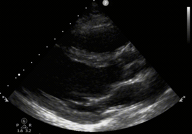
Right Ventricule Evaluation
The right ventricle is crescent shape that wraps along the LV. This adds complexity to the quantification of its size and function
Our focus here on cardiac ultrasound is to be able to determine RV size and function in a qualitative or semiquantitative way in an interrogation that is conducted without the requirement of performing specific measurements. We will explore some qualitative appreciation of sizes and function of the RV and go beyond the scope of FoCUS to be give you numerical data you can use clinically.
1. Visual Global Assessment
Comparing right ventricular dimensions to left ventricle dimensions is an important technique to assess function. The ideal views for this technique are apical 4-chamber and subcostal 4-chamber views. The RV internal diameter should not be more than 2/3 the size of the LV, and it should not extend more than 2/3 to the apex of the LV. Changes in the size of the RV as well as displacement of the tricuspid valve can be used to evaluate function. Linear measurements are very useful to determine function (see later section)
Normal RV Dimensions and Function
-RV internal diameter should be <2/3 of the size of the LV
-RV should extend <2/3 to the apex of the LV
-The interventricular septum should not be flattened or D shaped which is indicative of RV pressure or volume overload
-The Systolic eccentricity index (EI) is a measure of RV overload. At end systole D1 bisects pap muscles and D2 is orthogonal. EI is D2/D1. Normal EI is 1 and dyskinesia is present when EI>1


D2
D1
Dilated and
Depressed RV
-Enlarged RV. The RV is bigger in size than the LV
-Depressed RV
-On systole and diastole the septum is pushed towards the LV indicating fluid and pressure overload.
-Enlarged RA


Comparison of RV Depression
-On the left the RV diameter does not appear enlarged and function seems mildly depressed.
-On the right, the RV is enlarged and depressed with both fluid and pressure overload. The free wall of the RV does not appear to move but the apex does (McConnell's sign)


2. Linear Dimensions of Size. Beyond FoCUS
Measurements by 2D ultrasound are challenging because of the complex geometry of the right ventricle and the lack of specific right-sided anatomic landmarks to be used as reference points. The apical four chamber view results in considerable variability in how the right heart is sampled. RV linear dimensions derived from these areas may vary widely in the same patient with relatively minor rotations in transducer position. RV measurements may also be limited when the RV free wall is not well defined because of the dimension of the ventricle itself or its position behind the sternum. In general, a diameter >41 mm at the base and >35 mm at the mid level in the apical 4 chamber view will indicate RV dilatation.


<41
<35
<27
<35
1
2
3
Linear dimension of the RV. The images above display the maximum measurements considered normal (in mm). 1, Modified 4 chamber view. A solid white shows the mid right ventricular diameter and the solid red line is at the base of the RV. 2, Inflow outflow view and out of the scope of the Focus exam views and showing the RVOT distal diameter. 3, Long axis view showing a proximal RVOT measurement.
Basal RV dimensions on the left image is the maximum dimensions obtained at the base of the RV inflow (red line). The mid cavitary RV linear dimensions taken at mid pap level with the white line. Both measurements on diastole. On the middle and right image the white dotted line is the proximal RV outflow diameter; taken from the anterior RV wall to the interventricular septal-aortic junction(right image) or the aortic valve (center image). Upper limit of normal displayed on images (measurements in mm).

<5
On the left, a 4 chamber subcostal view. Linear thickness of the RV free wall and end diastole with pap muscles excluded. Upper limit of normal (in mm) displayed on the image. The best view to measure the RV thickness in on the subcostal window.
3. Linear Dimensions to Estimate RV EF. Beyond FoCUS
There are multiple numeric parameters to evaluate RV function. We will take a look at the easiest to perform.
3A. Fractional Area Change RV
FAC provides an estimate of global RV systolic function. The apex and free wall must be included in the measurement. RV FAC < 35% indicates RV systolic dysfunction. In the images below, from left to right end diastole and systole.


/
/
/
/
/
/
|
|
_
_
_
_
_
_
|
|
|
|
\
\
\
|
FAC is calculated as FAC=(End Diastolic Area - End Systolic Area)/ End Diastolic Area
3B.TAPSE
Trans Annular Plane Excursion or TAPSI represents a measure of the RV longitudinal function and measured using M-mode. The cursor should have proper alignment on the apical 4 chamber view and along the direction of the tricuspid lateral annulus. The drawbacks is that it depends on angle of measurement and is only partially representative of RV global function. A value less than 17mm is highly suggestive of RV systolic dysfunction.


On the left, the cursor is aligned with the tricuspid annulus. On the right a TAPSI measurement indicated with the red dots. It a measure of longitudinal displacement.
References
1. Lang RM, Badano LP, Mor-Avi V, et al. Recommendations for cardiac chamber quantification by echocardiography in adults: an update from the American Society of Echocardiography and the European Association of Cardiovascular Imaging. J Am Soc Echocardiogr. 2015;28(1):1-39.e14.
2. Via G, Hussain A, Wells M, Reardon R, ElBarbary M, Noble VE, Tsung JW, Neskovic AN, Price S, Oren-Grinberg A, Liteplo A, Cordioli R, Naqvi N, Rola P, Poelaert J, Guliĉ TG, Sloth E, Labovitz A, Kimura B, Breitkreutz R, Masani N, Bowra J, Talmor D, Guarracino F, Goudie A, Xiaoting W, Chawla R, Galderisi M, Blaivas M, Petrovic T, Storti E, Neri L, Melniker L; International Liaison Committee on Focused Cardiac UltraSound (ILC-FoCUS); International Conference on Focused Cardiac UltraSound (IC-FoCUS). International evidence-based recommendations for focused cardiac ultrasound. J Am Soc Echocardiogr. 2014 Jul;27(7):683.e1-683.e33. doi: 10.1016/j.echo.2014.05.001. PMID: 24951446.
3. Gudmundsson P, Rydberg E, Winter R, Willenheimer R. Visually estimated left ventricular ejection fraction by echocardiography is closely correlated with formal quantitative methods. International Journal of Cardiology,
2005; Vol 101: Issue 2: 209-212. ISSN 0167-5273.


